


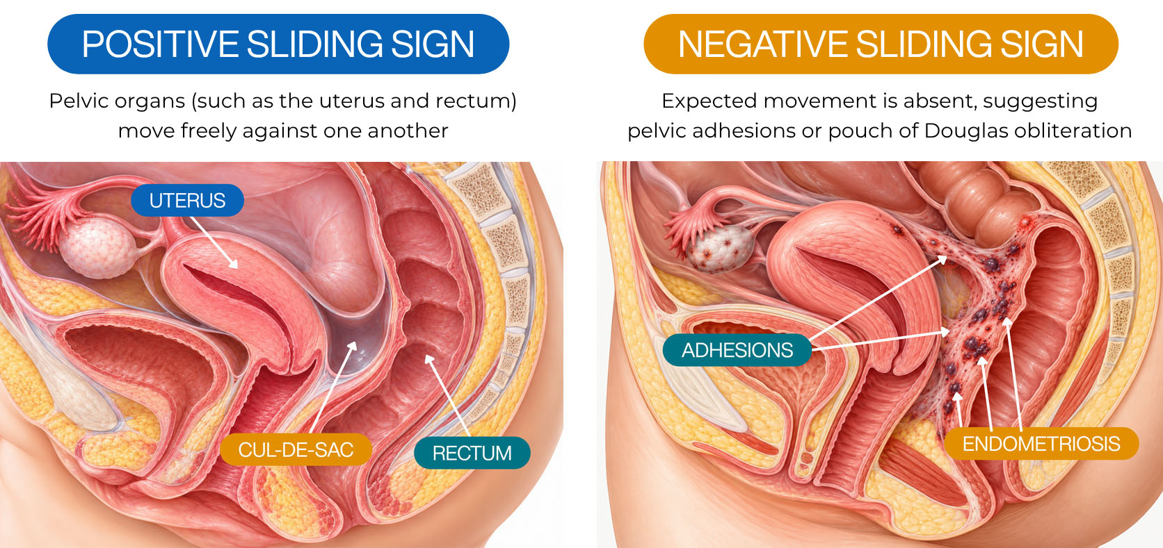
A negative sliding sign on ultrasound means that the expected movement between certain pelvic structures is absent during a dynamic ultrasound examination. This finding can suggest pelvic adhesions or pouch of Douglas obliteration, particularly when restricted movement is detected between the uterus and bowel. [1][3]
In patients being evaluated for endometriosis, a negative sliding sign may also raise suspicion for associated deep endometriosis. However, the finding does not establish the presence, cause, or exact extent of the disease.
A negative sliding sign is an important imaging finding, but it is not a diagnosis of endometriosis. Understanding what it reveals, and what it cannot determine, is essential when interpreting an ultrasound report.
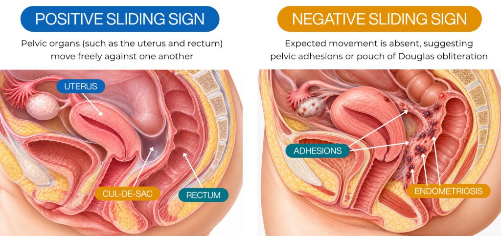
Medical illustration comparing positive and negative sliding signs. A positive sliding sign demonstrates normal movement between the uterus and rectum, while a negative sliding sign shows restricted mobility associated with pelvic adhesions and possible pouch of Douglas obliteration.
What Is the Sliding Sign on Ultrasound?
The sliding sign is a dynamic ultrasound assessment used to determine whether adjacent pelvic structures move freely against one another.
Unlike conventional ultrasound images, which primarily show anatomical structures, the sliding sign evaluates their movement during an examination.
During transvaginal ultrasound, the examiner uses gentle probe pressure and, when appropriate, abdominal pressure to assess movement between the bowel and uterus. [3]
The exact technique depends on the position of the uterus.
The results are generally interpreted as follows:
- Positive sliding sign: The examined structures move freely against one another at the assessed location.
- Negative sliding sign: The expected sliding movement is absent, suggesting restricted mobility or possible adhesions.
- Equivocal sliding sign: Movement cannot be confidently interpreted, potentially requiring further assessment.
A technically inadequate examination should not be interpreted as a negative sliding sign.
The sliding sign is particularly useful when evaluating the posterior pelvic compartment in patients with suspected endometriosis. [3][6]
A Negative Sliding Sign May Indicate Pelvic Adhesions
Pelvic adhesions are bands of fibrous tissue that connect structures that would otherwise move more independently.
These adhesions can develop after surgery, infection, inflammation, or endometriosis. When they connect nearby organs, normal movement may become restricted. [3]
For example, adhesions may limit movement between the uterus and rectum or contribute to an ovary becoming fixed near surrounding structures.
However, not all pelvic adhesions are caused by endometriosis.
A negative sliding sign identifies a possible mechanical restriction but does not independently determine whether its underlying cause is endometriosis, previous surgery, or another condition.
Pouch of Douglas Obliteration Is an Important Finding
The pouch of Douglas, also called the rectouterine pouch or posterior cul-de-sac, is the peritoneal space between the uterus and rectum.
In normal pelvic anatomy, adjacent surfaces generally move relative to one another.
When adhesions or fibrosis cause these structures to become attached, the pouch of Douglas may become partially or completely obliterated.
During a specialized ultrasound examination, the clinician evaluates whether the bowel moves freely against the posterior uterus.
For an anteverted uterus, the assessment includes movement between the anterior rectal wall and retrocervix, as well as between the anterior sigmoid wall and posterior uterine fundus. A retroverted uterus requires a modified technique. [3]
Absence of the expected sliding movement at an assessed posterior interface may indicate pouch of Douglas obliteration. [3]
In a 2013 prospective study involving 100 women undergoing laparoscopy for suspected endometriosis, Reid and colleagues reported that the sliding sign had 83.3% sensitivity and 97.1% specificity for predicting pouch of Douglas obliteration. [5]
These findings support its value in preoperative assessment, although imaging findings must be interpreted within the broader clinical picture.
The Sliding Sign Can Help Identify Deep Endometriosis
Deep endometriosis can affect structures such as the uterosacral ligaments, rectovaginal region, and bowel.
Associated fibrosis and adhesions may alter normal anatomical relationships and restrict tissue mobility.
A negative sliding sign can therefore raise concern for more extensive pelvic disease, particularly when accompanied by other suspicious ultrasound findings. [3][6]
These may include:
- Ovarian endometriomas
- Ovaries with restricted mobility
- Deep endometriotic nodules
- Abnormalities involving the uterosacral ligaments
- Suspicious lesions affecting the rectum or rectosigmoid region
However, these findings should be evaluated individually.
An ovarian endometrioma does not, by itself, establish deep endometriosis, and a negative sliding sign cannot identify every lesion or determine the full extent of pelvic disease. [2][3]
A Negative Sliding Sign Does Not Confirm Bowel Endometriosis
Bowel endometriosis and bowel adhesions are related but distinct findings.
A bowel segment may be restricted by adhesions involving its outer surface without endometriotic tissue infiltrating the bowel wall. [1][3]
Conversely, endometriotic lesions may involve the bowel even when the sliding sign does not demonstrate the expected abnormality.
Therefore, a negative sliding sign should not be interpreted as direct evidence of bowel-wall infiltration.
When bowel endometriosis is suspected, a targeted ultrasound examination or pelvic MRI may help characterize its location and extent. [3][4]
This distinction is particularly important when planning surgery.
How Accurate Is the Sliding Sign?
A 2022 systematic review and meta-analysis by Alcázar and colleagues assessed the diagnostic accuracy of the sliding sign in patients with suspected endometriosis.
Eight studies were included, with 938 patients contributing to the analysis of pouch of Douglas obliteration and 963 to the analysis of bowel involvement.
The researchers compared ultrasound findings with laparoscopic findings. [1]
| Condition Assessed | Sensitivity | Specificity |
|---|---|---|
| Pouch of Douglas obliteration | 88% | 94% |
| Bowel involvement | 81% | 95% |
Source: Alcázar et al., 2022. [1]
Sensitivity describes how often a test correctly identifies patients who have the condition.
Specificity describes how often a test correctly identifies patients who do not have the condition.
These findings demonstrate that the sliding sign can be useful in specialized endometriosis assessment.
However, the review reported substantial variation between studies evaluating bowel involvement. Diagnostic performance may also differ according to examiner expertise, examination technique, and patient characteristics. [1]
Importantly, these percentages do not represent an individual patient’s probability of having endometriosis.
The sliding sign primarily evaluates restricted mobility and related anatomical findings rather than detecting all forms of the disease.
A Positive Sliding Sign Does Not Exclude Endometriosis
A positive sliding sign indicates that the examined structures demonstrate the expected movement at the assessed location.
Although reassuring regarding mobility, it does not establish that the entire pelvis is free from adhesions or endometriosis.
Superficial peritoneal endometriosis may occur without substantial fibrosis or restricted organ movement. Lesions may also exist in pelvic locations not directly assessed by the sliding-sign maneuver.
Additionally, incomplete posterior compartment obliteration may sometimes be present despite apparently preserved sliding.
The European Society of Human Reproduction and Embryology (ESHRE) emphasizes that negative ultrasound or MRI findings cannot exclude endometriosis, particularly superficial peritoneal disease. [2]
Therefore, persistent pelvic pain, painful menstruation, painful intercourse, bowel symptoms associated with menstruation, or fertility concerns may warrant further evaluation even when ultrasound findings appear reassuring.
Why a Negative Sliding Sign Matters Before Surgery
A negative sliding sign may help surgeons anticipate pelvic adhesions, distorted anatomy, or pouch of Douglas obliteration before an operation. [3]
These findings can guide surgical planning, particularly when bowel involvement is suspected. [3][4]
However, a negative sliding sign does not automatically mean surgery is necessary. Treatment depends on symptoms, imaging findings, fertility goals, and individual circumstances. [2][4]
What Should You Do If Your Ultrasound Shows a Negative Sliding Sign?
A negative sliding sign should be interpreted alongside symptoms, medical history, and other imaging findings.
Depending on the clinical situation, an endometriosis specialist may recommend specialized transvaginal ultrasound or pelvic MRI to assess possible adhesions or deep endometriosis. [4]
Diagnostic laparoscopy may also be considered when endometriosis is suspected, even if other imaging is normal. The decision depends on symptoms, prior treatment, fertility goals, and individual clinical findings. [2][4]

Can the NYGSE Endometriosis Likelihood Index Help?
The NYGSE Endometriosis Likelihood Index (ELI) is an educational tool that estimates the likelihood of endometriosis using selected clinical factors.
It may help patients organize information relevant to a discussion with an endometriosis specialist.
However, the ELI is not a diagnostic tool; it only estimates likelihood. It cannot confirm adhesions, determine whether the pouch of Douglas is obliterated, or establish the presence of deep bowel endometriosis.
A negative sliding sign requires independent clinical interpretation and should not be considered diagnostic based on an ELI estimate.

Understanding Your Ultrasound Findings
A negative sliding sign on ultrasound suggests restricted pelvic organ movement, which may indicate adhesions or pouch of Douglas obliteration. However, it does not independently confirm endometriosis or determine the extent of disease.
At New York Gynecology Surgery & Endometriosis (NYGSE), ultrasound findings are evaluated alongside symptoms and medical history to guide appropriate diagnosis and individualized treatment.
This article is for general education and isn’t medical advice for any individual. If you have sudden severe pelvic pain, heavy bleeding, fever or fainting, seek urgent care.
References
- Alcázar, J. L., Eguez, P. M., Forcada, P., Ternero, E., Martínez, C., Pascual, M. Á., & Guerriero, S. (2022). Diagnostic accuracy of sliding sign for detecting pouch of Douglas obliteration and bowel involvement in women with suspected endometriosis: Systematic review and meta-analysis. Ultrasound in Obstetrics & Gynecology, 60(4), 477–486. https://doi.org/10.1002/uog.24900
- Becker, C. M., Bokor, A., Heikinheimo, O., Horne, A., Jansen, F., Kiesel, L., King, K., Kvaskoff, M., Nap, A., Petersen, K., Saridogan, E., Tomassetti, C., van Hanegem, N., Vulliemoz, N., & Vermeulen, N. (2022). ESHRE guideline: Endometriosis. Human Reproduction Open, 2022(2), hoac009. https://doi.org/10.1093/hropen/hoac009
- Guerriero, S., Condous, G., van den Bosch, T., Valentin, L., Leone, F. P. G., van Schoubroeck, D., Exacoustos, C., Installé, A. J. F., Martins, W. P., Abrão, M. S., Hudelist, G., Bazot, M., Alcázar, J. L., Gonçalves, M. O., Pascual, M. A., Ajossa, S., Savelli, L., Dunham, R., Reid, S., Timmerman, D. (2016). Systematic approach to sonographic evaluation of the pelvis in women with suspected endometriosis, including terms, definitions and measurements: A consensus opinion from the International Deep Endometriosis Analysis (IDEA) group. Ultrasound in Obstetrics & Gynecology, 48(3), 318–332. https://doi.org/10.1002/uog.15955
- National Institute for Health and Care Excellence. (2024). Endometriosis: Diagnosis and management (NICE Guideline No. NG73). https://www.nice.org.uk/guidance/ng73
- Reid, S., Lu, C., Casikar, I., Reid, G., Abbott, J., Cario, G., Chou, D., Kowalski, D., Cooper, M., & Condous, G. (2013). Prediction of pouch of Douglas obliteration in women with suspected endometriosis using a new real-time dynamic transvaginal ultrasound technique: The sliding sign. Ultrasound in Obstetrics & Gynecology, 41(6), 685–691. https://doi.org/10.1002/uog.12305
- Young, S. W., Jha, P., Chamié, L., Rodgers, S., Kho, R. M., Horrow, M. M., Glanc, P., Feldman, M., Groszmann, Y., Khan, Z., Young, S. L., Poder, L., Burnett, T. L., Hu, E. M., Egan, S., & VanBuren, W. (2024). Society of Radiologists in Ultrasound consensus on routine pelvic US for endometriosis. Radiology, 311(1), e232191. https://doi.org/10.1148/radiol.232191