Seeing “uterosacral ligament thickening” or “uterosacral ligament nodularity” in an MRI report can be confusing. These ligaments are located deep within the pelvis and help support the uterus and cervix.
They are also an important location for deep endometriosis. When an MRI shows abnormal thickening, nodularity, or irregularity of a uterosacral ligament, endometriosis may be one possible explanation, particularly when other symptoms or imaging findings support the diagnosis. [1][2]
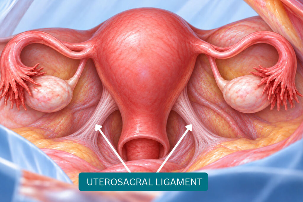
Medical illustration of normal uterosacral ligaments
Understanding the Uterosacral Ligaments
The uterosacral ligaments extend from the region around the cervix toward the sacrum at the back of the pelvis.
These ligaments are part of the support system of the female pelvis. They help support the cervix and uterus and contribute to maintaining their position within the pelvis. Their location also places them close to several important pelvic structures.
Because of their location behind the uterus, endometriosis involving these ligaments can occur alongside disease affecting nearby structures, including the posterior cervix, pouch of Douglas, vagina, rectum, or surrounding pelvic tissues. [2]
This anatomy is one reason the uterosacral ligaments are an important area to evaluate when deep endometriosis is suspected.
Why Does Endometriosis Affect These Ligaments?
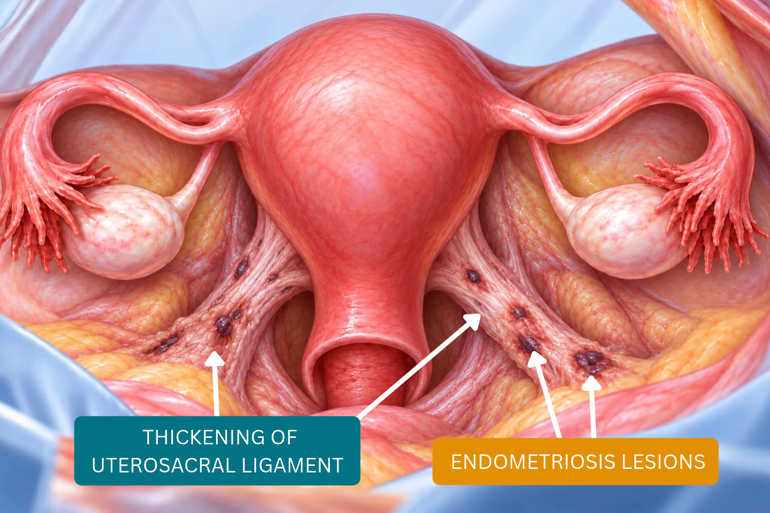
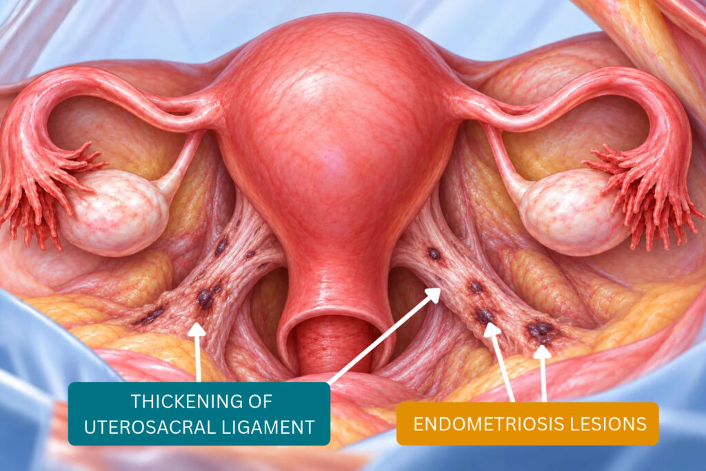
Deep endometriosis can produce fibrosis and nodular tissue within or around the uterosacral ligaments. [1][2]
On MRI, this may appear as thickening, a nodule, spiculation, or an irregular fibrotic area. Current European Society of Urogenital Radiology (ESUR) recommendations consider a uterosacral ligament abnormal when a nodule or spiculation is visible in at least two imaging planes or when a characteristic bright T1-weighted focus is present. Thickening alone is less specific for endometriosis. [1][2]
Medical illustration comparing normal uterosacral ligaments with thickened or nodular ligaments associated with deep endometriosis.
What Does Uterosacral Ligament Endometriosis Look Like on MRI?
MRI may show one ligament looking thicker or more irregular than the other. A radiologist may describe findings such as:
Uterosacral ligament thickening
Nodular thickening
Fibrotic thickening
Spiculation
A discrete uterosacral ligament nodule
The location can also matter. MRI reports may specify whether the finding involves the right or left ligament and whether it is closer to the cervix or extends farther into the posterior pelvis. [1][2]
Does Thickening Always Mean Endometriosis?
No.
Uterosacral ligament thickening by itself does not confirm endometriosis. Imaging findings need to be interpreted together with symptoms, examination findings, other areas seen on MRI or ultrasound, and the patient’s medical history. [1][2]
This distinction is important because even expert MRI criteria do not consider ligament thickness alone specific enough to diagnose an endometriotic deposit. [1][2]
Symptoms Associated With Uterosacral Ligament Endometriosis
Some patients may have no symptom that points specifically to the uterosacral ligaments.
When symptoms are present, they may occur as part of a broader pattern of deep endometriosis and can include pelvic pain, painful periods, deep pain during sex, or pain that extends toward the lower back or posterior pelvis. [4]
Symptoms alone cannot determine exactly where endometriosis is located.
Can MRI Help Before Endometriosis Surgery?
Yes. MRI can be particularly useful for mapping deep endometriosis before surgery.[3]
Knowing that disease may involve the uterosacral ligaments and nearby structures can help the surgical team understand the distribution of suspected disease and plan an appropriate approach. Current ESUR recommendations specifically support MRI in patients being considered for surgical or interventional treatment of endometriosis.
When Uterosacral Ligament Thickening Deserves Further Evaluation
A finding of uterosacral ligament thickening or nodularity does not automatically mean that surgery is necessary.
However, when it appears alongside persistent pelvic pain, painful periods, deep pain during sex, endometriomas, bowel symptoms, or other MRI findings associated with deep endometriosis, further evaluation may be appropriate.
At New York Gynecology Surgery & Endometriosis (NYGSE), we consider the complete pattern of symptoms and imaging findings to determine whether deep endometriosis may be contributing and whether medical management, further imaging, surgical evaluation, or another approach may be appropriate.
Becker, C. M., Bokor, A., Heikinheimo, O., Horne, A., Jansen, F., Kiesel, L., King, K., Kvaskoff, M., Nap, A., Petersen, K., Saridogan, E., Tomassetti, C., van Hanegem, N., Vulliemoz, N., & Vermeulen, N. (2022). ESHRE guideline: Endometriosis. Human Reproduction Open, 2022(2), hoac009. https://doi.org/10.1093/hropen/hoac009
Stoppa, M., et al. (2023). Mild endometriosis of the uterosacral ligaments: A retrospective study of magnetic resonance imaging performance for diagnosis. Reproductive BioMedicine Online, 46(6), 947–955. https://doi.org/10.1016/j.rbmo.2023.03.002
Thomassin-Naggara, I., et al. (2025). ESUR consensus MRI for endometriosis: Indications, reporting, and classifications. European Radiology, 35(11), 7260–7268. https://doi.org/10.1007/s00330-025-11579-0
Thomassin-Naggara, I., et al. (2025). ESUR consensus MRI for endometriosis: Protocol, lexicon, and compartment-based analysis. European Radiology, 35(11), 7272–7286. https://doi.org/10.1007/s00330-025-11611-3