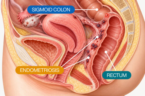
Hearing that there is a “blood clot in the ovary” can be confusing. The phrase may sound like a clot is growing inside the ovary, but medically, several different conditions can involve blood or clot-like material in or around an ovarian structure.
One possibility is an ovarian endometrioma, a cyst associated with endometriosis that contains blood products from repeated bleeding. Another is a hemorrhagic ovarian cyst, which forms when bleeding occurs within a functional ovarian cyst. A very different condition, called ovarian vein thrombosis, involves a blood clot in the vein that drains the ovary rather than inside the ovary itself. [4]
Endometriomas Can Contain Old Blood and Blood Clots
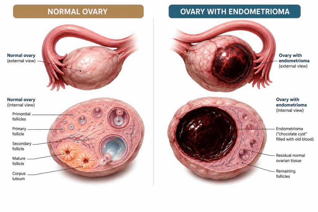
Because bleeding can occur repeatedly over time, the contents of an endometrioma may include blood products of different ages. This chronic hemorrhage contributes to the characteristic appearance of endometriomas on imaging. [3]
This is also why endometriomas are sometimes informally called “chocolate cysts”. The term refers to their thick, old blood content rather than fresh bleeding.
Importantly, an endometrioma is not the same thing as a conventional blood clot circulating through a blood vessel.
Blood Clots Can Be Seen Within an Endometrioma
Research has identified imaging findings that may represent chronic retractile blood clots within endometriomas.
One example is the T2 dark spot sign on MRI. These dark spots are thought to represent chronic retracted blood clots within an endometrioma. Corwin et al. (2014) found that the sign had 36% sensitivity and 93% specificity for distinguishing endometriomas from other hemorrhagic ovarian lesions. [2]
The presence of a clot-like area inside an ovarian cyst therefore does not necessarily mean that a patient has a dangerous vascular blood clot.
Medical illustration comparing a normal ovary with an ovarian endometrioma
Endometrioma vs. Hemorrhagic Ovarian Cyst
A hemorrhagic ovarian cyst can also contain blood and retracting clots.
These cysts usually develop when bleeding occurs within a functional ovarian cyst. On ultrasound, fibrin strands and retracting clots may be visible. Because both hemorrhagic cysts and endometriomas contain blood products, their appearances can sometimes overlap. [4]
MRI can provide additional information when ultrasound findings are uncertain. Endometriomas often demonstrate characteristic findings related to chronic blood products, including T2 shading, while other MRI characteristics can help distinguish them from hemorrhagic cysts. [4]
The distinction matters because an endometrioma represents ovarian endometriosis, whereas a hemorrhagic functional cyst is a different ovarian condition.
Ovarian Vein Thrombosis Is Different
An ovarian vein thrombosis (OVT) is a true vascular blood clot that develops in an ovarian vein rather than within an endometrioma or ovarian cyst. [5]
OVT is uncommon and belongs to the spectrum of venous thromboembolism. It has been reported particularly in association with pregnancy and the postpartum period, malignancy, pelvic infection, recent surgery, and other thrombotic risk factors. [5]
This distinction is important:
Endometrioma: blood products accumulate within an ovarian cyst associated with endometriosis.
Hemorrhagic ovarian cyst: bleeding occurs within an ovarian cyst and may produce a retracting clot.
Ovarian vein thrombosis: a thrombus forms within the vein that drains the ovary.
These conditions should not be described interchangeably.
What a “Blood Clot in the Ovary” May Mean
If imaging, surgery, or a medical report mentions blood or a clot involving an ovary, the exact terminology matters.
At New York Gynecology Surgery & Endometriosis (NYGSE), Dr. Pankaj Singhal and Dr. Elizabeth McLean evaluate ovarian findings as part of the broader assessment for endometriosis and other gynecologic conditions.
Understanding whether a finding represents an endometrioma, hemorrhagic ovarian cyst, or a vascular blood clot is an important first step in determining what it means and what evaluation or treatment may be appropriate.
Becker, C. M., Bokor, A., Heikinheimo, O., Horne, A., Jansen, F., Kiesel, L., King, K., Kvaskoff, M., Nap, A., Petersen, K., Saridogan, E., Tomassetti, C., van Hanegem, N., Vulliemoz, N., & Vermeulen, N. (2022). ESHRE guideline: Endometriosis. Human Reproduction Open, 2022(2), hoac009. https://doi.org/10.1093/hropen/hoac009
Corwin, M. T., Gerscovich, E. O., Lamba, R., Wilson, M., & McGahan, J. P. (2014). Differentiation of ovarian endometriomas from hemorrhagic cysts at MR imaging: Utility of the T2 dark spot sign. Radiology, 271(1), 126–132. https://doi.org/10.1148/radiol.13131394
Decommer, K., & Feyaerts, F. (2020). The T2 dark spot sign in endometrioma. Journal of the Belgian Society of Radiology, 104(1), 74. https://doi.org/10.5334/jbsr.2271
Levine, D., et al. (2010). Management of Asymptomatic Ovarian and Other Adnexal Cysts Imaged at US Consensus Conference Statement Panel. (2010). Management of asymptomatic ovarian and other adnexal cysts imaged at US: Society of Radiologists in Ultrasound consensus conference statement. Radiology, 256(3), 943–954. https://doi.org/10.1148/radiol.10100213
Riva, N., & Calleja-Agius, J. (2021). Ovarian vein thrombosis: A narrative review. Hämostaseologie, 41(4), 257–266. https://doi.org/10.1055/a-1306-4327