


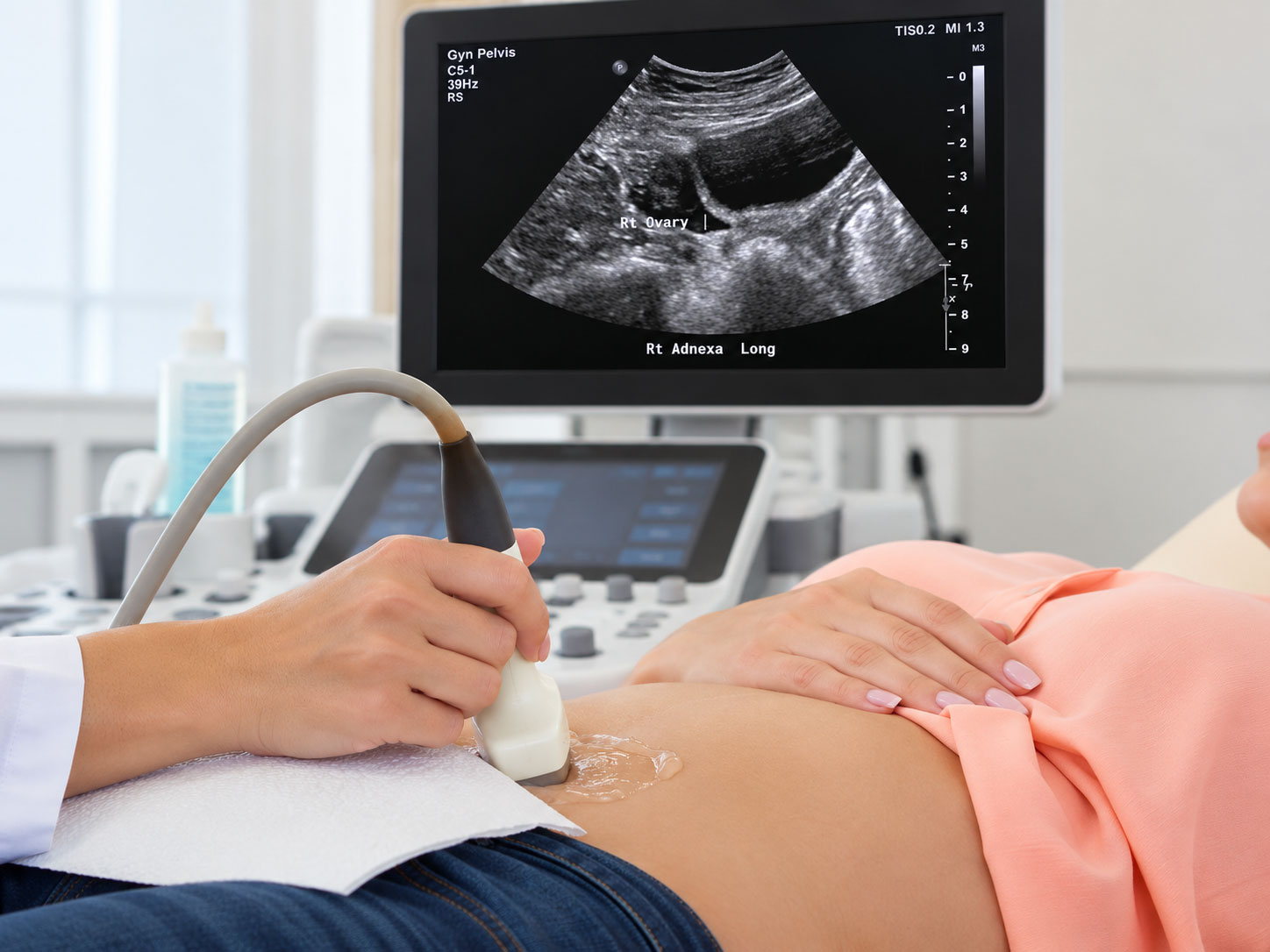
You have painful periods. Sometimes the pain continues long after your period ends. Your doctor orders a pelvic ultrasound, but the results come back normal.
Nothing unusual was found.
Does that mean nothing is wrong?
Not necessarily. Endometriosis can be present even when a pelvic ultrasound appears completely normal. Some forms of the disease are visible on imaging, while others may remain undetected. [1][2]
The important question is not simply whether your ultrasound is normal, but what the examination was capable of detecting.
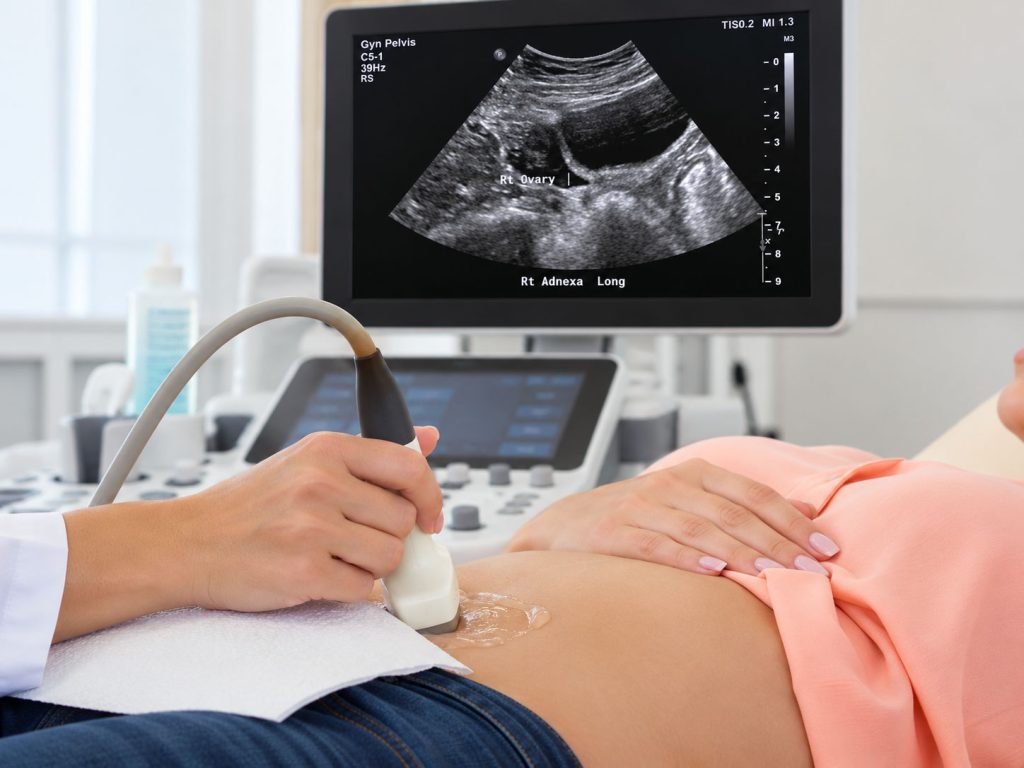
Woman undergoing a pelvic ultrasound examination while a monitor displays an ultrasound image.
A Normal Ultrasound Does Not Rule Out Endometriosis
Ultrasound uses sound waves to create images of structures inside the pelvis, particularly the uterus and ovaries. It can identify ovarian cysts, uterine fibroids, and certain abnormalities that may explain pelvic pain.
But endometriosis does not always produce visible changes.
Endometriosis occurs when tissue resembling the lining of the uterus grows outside the uterus. These lesions may develop on the ovaries, pelvic lining, supporting ligaments, and surrounding structures.
Some lesions form recognizable cysts or nodules. Others remain small, flat, or difficult to distinguish from normal tissue.
An ultrasound can show normal pelvic anatomy without revealing every endometriosis lesion that may be present. [1][2]
This does not necessarily mean the examination was performed incorrectly. It reflects the limitations of the imaging technique.
Why Doesn't Endometriosis Always Show Up on Ultrasound?
Not all endometriosis looks or behaves the same way.
Some forms create abnormalities that ultrasound can identify. Others involve thin areas of diseased tissue without substantially changing the appearance of pelvic organs.
Understanding these differences helps explain why ultrasound detects endometriosis in some patients but misses it in others.
Superficial Endometriosis Is Difficult to Detect
The pelvis is lined by a thin membrane called the peritoneum. It covers portions of the pelvic cavity and provides a surface over which pelvic organs can move.
Superficial peritoneal endometriosis develops on this membrane. These lesions may appear as small spots or patches rather than distinct masses.
Think of a small stain on a wall. Equipment designed to examine the structure behind the wall would not necessarily reveal what is happening on its surface.
Ultrasound faces a similar limitation.
Superficial endometriosis may be present even when the uterus and ovaries appear completely normal. This is why a normal ultrasound cannot reliably exclude this form of the disease. [1][3]
Deep Endometriosis May Require Specialized Imaging
Deep endometriosis extends beneath the pelvic surface and may involve structures near the bowel, bladder, or uterosacral ligaments.
Unlike superficial lesions, deep endometriosis can produce nodules, tissue thickening, or changes in how pelvic organs move.
These abnormalities may be detected through specialized transvaginal ultrasound performed by clinicians experienced in identifying endometriosis.
Ovarian endometriomas, cysts associated with endometriosis, are also often visible on ultrasound.
However, a routine pelvic ultrasound is not necessarily equivalent to a specialized examination for deep endometriosis. The examination technique and experience of the clinician can influence what is detected. [2][3]
Can You Have Severe Pelvic Pain With a Normal Ultrasound?
Yes. And this raises an important question.
If the ultrasound looks normal, where is the pain coming from?
Ultrasound shows anatomical structures. It does not measure pain.
Endometriosis can contribute to discomfort through inflammation, irritation of nearby nerves, and changes in how the nervous system processes painful signals.
Importantly, the amount of endometriosis does not always correspond to the severity of symptoms.
A patient with relatively limited disease may experience substantial pain, while another with more extensive endometriosis may report fewer symptoms. [1]
However, endometriosis is not the only possible explanation.
Adenomyosis, pelvic floor muscle dysfunction, bladder conditions, and gastrointestinal disorders can also cause pelvic pain.
Persistent symptoms therefore deserve evaluation rather than an assumption that normal imaging means nothing is wrong.
What Should You Do If Your Ultrasound Is Normal?
A normal ultrasound provides useful information, but it should be considered alongside your symptoms and medical history.
Does your pain worsen during menstruation? Is intercourse painful? Do bowel movements or urination cause discomfort?
These details may help identify patterns that imaging alone cannot explain.
If you are unsure whether your symptoms could be associated with endometriosis, NYGSE’s Endometriosis Symptom Checker can help you explore common symptoms and prepare for a more informed discussion with your healthcare provider. The checker is an educational tool, not a diagnostic test.
When to Consider MRI or Further Evaluation
MRI may help identify deep endometriosis, particularly when the disease could involve the bowel, bladder, or other pelvic structures. However, MRI also cannot reliably exclude superficial endometriosis. [1][3]
Depending on symptoms and clinical findings, treatment may sometimes begin without surgical confirmation.
In selected cases, laparoscopy may be considered. This minimally invasive surgical procedure allows direct examination of pelvic structures and, when appropriate, treatment of visible disease.
Not every patient with suspected endometriosis requires surgery. The decision depends on symptoms, previous treatments, imaging findings, and individual circumstances. [1][2]
When to Seek an Endometriosis Specialist
If pelvic pain continues despite normal imaging, further evaluation may be appropriate, particularly when symptoms interfere with daily activities or persist despite treatment. [2]
An endometriosis specialist can review previous imaging, evaluate symptoms, and determine whether additional investigations are necessary.
Sometimes specialized imaging is appropriate. In other cases, medical treatment or surgery may be considered.
The goal is not to prove that the original ultrasound was wrong. It is to determine whether the findings adequately explain your symptoms.
Your Symptoms Matter, Even When Imaging Is Normal
A normal ultrasound can provide valuable information about pelvic anatomy, but it cannot identify every form of endometriosis.
The absence of visible endometriosis is not proof that the disease is absent.
At New York Gynecology Surgery & Endometriosis, evaluating suspected endometriosis involves considering symptoms, medical history, imaging findings, and previous treatments.
If pelvic pain persists despite normal ultrasound results, consulting an experienced endometriosis specialist may help determine the appropriate next steps.
Your ultrasound may be normal. Your symptoms still deserve a careful explanation.
References
- Becker, C. M., Bokor, A., Heikinheimo, O., Horne, A., Jansen, F., Kiesel, L., King, K., Kvaskoff, M., Nap, A., Petersen, K., Saridogan, E., Tomassetti, C., van Hanegem, N., Vulliemoz, N., & Vermeulen, N. (2022). ESHRE guideline: Endometriosis. Human Reproduction Open, 2022(2), hoac009. https://doi.org/10.1093/hropen/hoac009
- National Institute for Health and Care Excellence. (2024). Endometriosis: Diagnosis and management (NICE Guideline NG73). https://www.nice.org.uk/guidance/ng73
- Nisenblat, V., Bossuyt, P. M. M., Farquhar, C., Johnson, N., & Hull, M. L. (2016). Imaging modalities for the non-invasive diagnosis of endometriosis. Cochrane Database of Systematic Reviews, 2016(2), CD009591. https://doi.org/10.1002/14651858.CD009591.pub2